I have developed a number of tissue-level mathematical models of the human retina in the healthy and the diseased eye. These models are formulated as systems of partial differential equations (PDEs) or ordinary differential equations (ODEs) and solved using both analytical methods (e.g. perturbation methods, stability and travelling wave analysis) and numerical methods (e.g. finite difference and finite element methods).
[11] Roberts, P. A.,2024. Mathematical Models of Retinal Oxygenation and Metabolism in Health and Disease. (invited review for Invest. Ophthalmol. Vis. Sci.) (under review, 60 pages)
Details to follow.
[10] Roberts, P.A., 2025. Perspective on running a mathematical and computational ophthalmology seminar series. Artif. Intell. Vis. Ophthalmol., 1(1):1-5. (invited opinion letter) DOI
Details to follow.
[9] Roberts, P.A., Thomas, C.N., Bellamy Plaice, G., Roberts, J.A., Jones, M.C., Andrews, J.W., Hill, L.J., 2025. Mathematical models of topically and intravitreally applied ranibizumab. Invest. Ophthalmol. Vis. Sci., 66(11):45 (26 pages). DOI (bioRxiv)
Details to follow.
[8] Tweedy, J.H., Dvoriashyna, M., Crawshaw, J.R., Overby, D.R., Repetto, R., Roberts, P.A., Spelman, T.A., Stewart, P.S., Foss, A.J.E., 2025. A Model of the Mechanisms Underpinning Unconventional Aqueous Humor Outflow. Invest. Ophthalmol. Vis. Sci., 66(4):75 (20 pages). DOI
Details to follow.
[7] Hernandez, R., Roberts, P.A., El-Bouri, W.K, 2023. Advancing Treatment of Retinal Disease through in silico Trials. Prog. Biomed. Eng., 5:022002 (41 pages) (invited review) DOI
An invited review of retinal modelling, considering the potential of mechanistic retinal models for use with in silico trials. The paper has a particular focus upon vascular and drug delivery models.
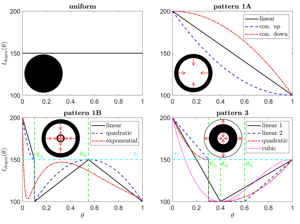
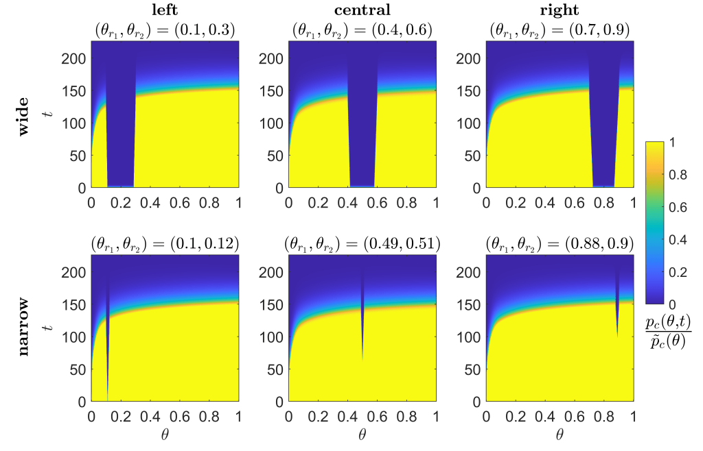
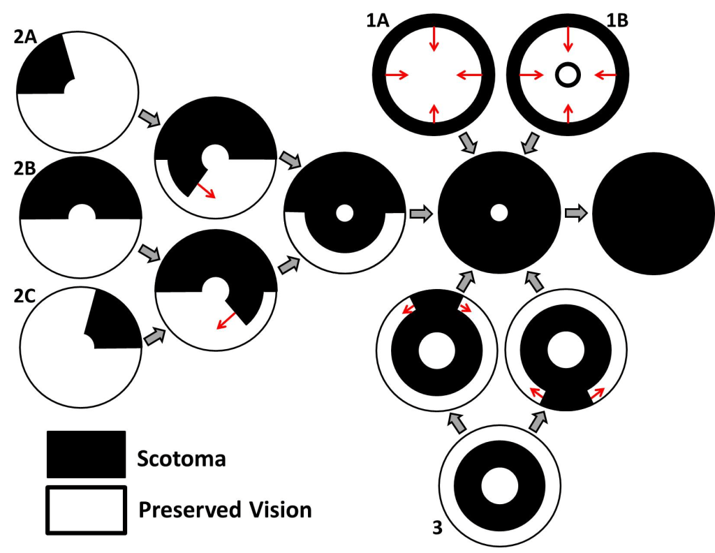
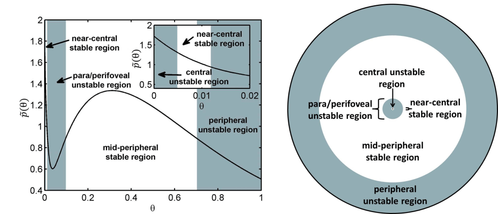
[6] Roberts, P.A., 2022. Inverse Problem Reveals Conditions for Characteristic Retinal Degeneration Patterns in Retinitis Pigmentosa under the Trophic Factor Hypothesis. Front. Aging Neurosci., 14(765966): (17 pages). (invited article) DOI (bioRxiv)
I derived and solved an inverse problem, revealing for the first time the experimentally testable conditions under which the trophic factor mechanism will qualitatively recapitulate the spatio-temporal patterns of retinal regeneration observed in the human form of the retinal degenerative disease retinitis pigmentosa.
[5] Roberts, P.A., 2022. Mathematical Models of Retinitis Pigmentosa: The Trophic Factor Hypothesis. J. Theor. Biol., 534:110938 (22 pages) (epub ahead of print, 2021). DOI (bioRxiv)
I developed the first spatial mathematical models of retinitis pigmentosa to explore the trophic factor hypothesis. These models answer the long-standing and much-debated question as to the patterns of retinal degeneration that would result if trophic factor loss was responsible for retinal degeneration. I also calculated the critical trophic factor treatment dosages required to prevent photoreceptor loss.
[4] Roberts, P.A., Gaffney, E.A., Whiteley, J.P., Luthert, P.J., Foss, A.J.E., Byrne, H.M., 2018. Predictive Mathematical Models for the Spread and Treatment of Hyperoxia-induced Photoreceptor Degeneration in Retinitis Pigmentosa. Invest. Ophthalmol. Vis. Sci., 59(3):1238-1249. DOI
I developed the first mathematical models of retinitis pigmentosa in two spatial dimensions. These models are the first to predict the patterns of degeneration that would result if oxygen toxicity was responsible for retinal degeneration, demonstrating the strengths and weaknesses of this hypothesis, where before there was only speculation. I also predicted the effects of treatment with antioxidants and trophic factors under a variety of clinically-relevant conditions.
[3] Roberts, P.A., Gaffney, E.A., Luthert, P.J., Foss, A.J.E., Byrne, H.M., 2017. Mathematical Models of Retinitis Pigmentosa: The Oxygen Toxicity Hypothesis. J. Theor. Biol., 425:53-71. DOI
I developed the first mathematical models of retinitis pigmentosa to account for the spatial distribution of photoreceptors or to consider oxygen toxicity as a disease mechanism. I determined the conditions under which patches of retinal degeneration will expand or remain stable, derived a formula for the speed of degeneration propagation and considered the effects of treatment with antioxidants and trophic factors.
[2] Roberts, P.A., Gaffney, E.A., Luthert, P.J., Foss, A.J.E., Byrne, H.M., 2016. Mathematical and Computational Models of the Retina in Health, Development and Disease. Prog. Retin. Eye Res., 53:48-69. (invited review) DOI
An invited review for the world-leading journal of ophthalmology. This paper gives a detailed account of the current state-of-the-art in mathematical and computational modelling of the retina and is the first review to have been written on this topic.
[1] Roberts, P.A., Gaffney, E.A., Luthert, P.J., Foss, A.J.E., Byrne, H.M., 2016. Retinal Oxygen Distribution and the Role of Neuroglobin. J. Math. Biol., 73(1):1-38. DOI
I discovered that the protein neuroglobin may prevent retinal oxygen starvation (hypoxia) and that, contrary to the prevailing view in the field, neuroglobin’s oxygen affinity is near optimal for oxygen transport. I also placed previous models of retinal oxygen distribution on a stronger theoretical foundation, demonstrating that they are valid provided oxygen levels do not become hypoxic, and showing how to modify the models in hypoxic regions.